Peer-reviewed and open access.

Nine papers on high-density peptide microarrays and their applications.

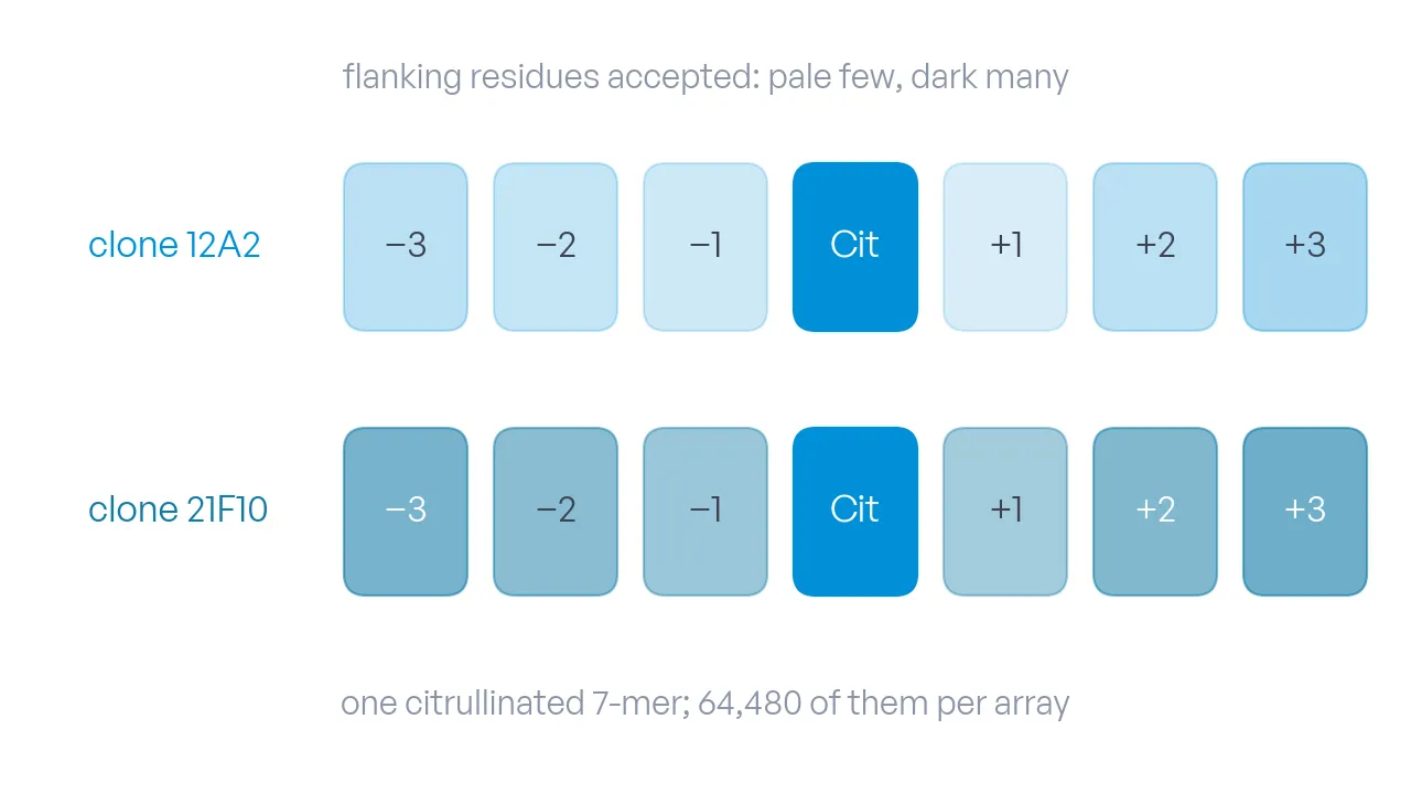
One citrullinated 7-mer as seven boxes, the citrulline in the middle; two rows for the two clones, the flanking positions shaded by how many residues each clone accepts.

2026 · Cell Reports Methods

DOI

Development of motif-specific monoclonal antibodies for global protein citrullination detection with minimal cross-reactivity to homocitrullination

Two antibody clones raised against a pool of over 490,000 citrullinated motif peptides are epitope-mapped on 64,480 citrullinated 7-mers in triplicate, which shows which flanking residues each clone accepts.
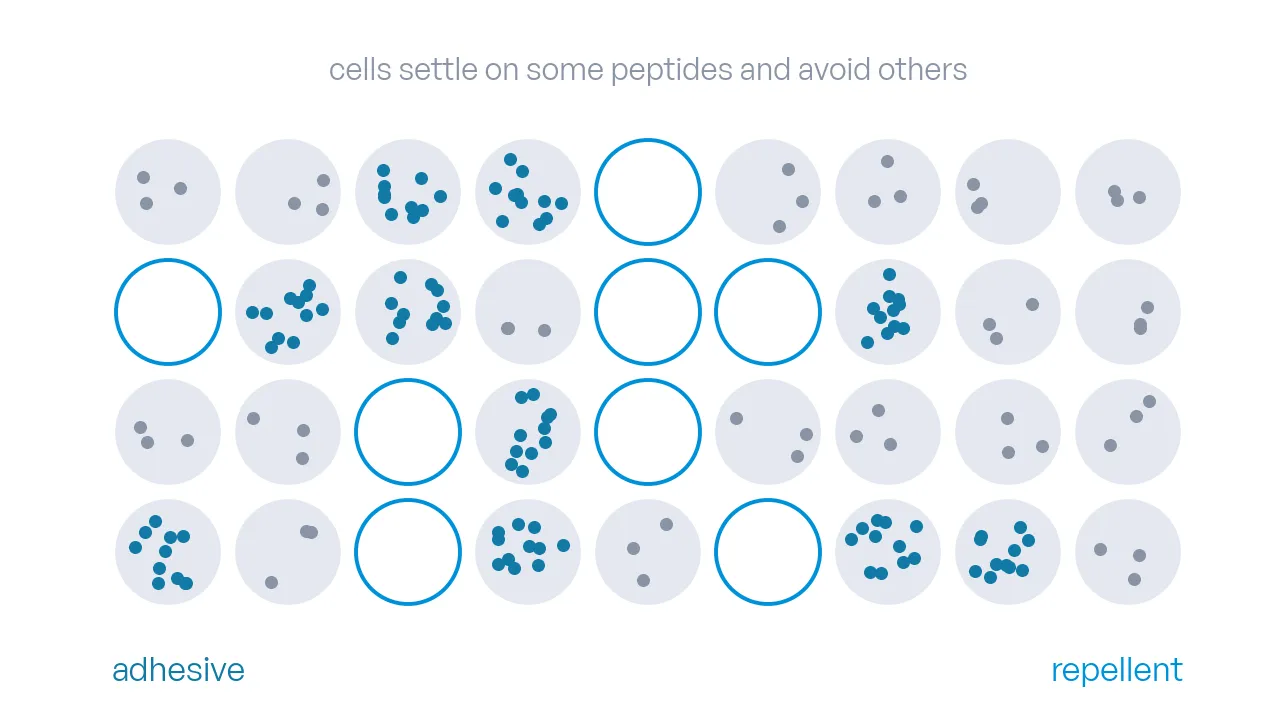
A grid of peptide spots: cells settled densely on adhesive spots, a few on most, none on the repellent spots drawn as empty rings.

2024 · Communications Biology

DOI

High-throughput screening for cell binding and repulsion peptides on multifunctionalized surfaces

Colorectal cancer cells grown on an array of over 11,000 peptides sort the library into strongly adhesive and strongly repellent sequences, among them repellent peptides from frizzled- and Dickkopf-related proteins and from the poles of the TNF-alpha trimer.

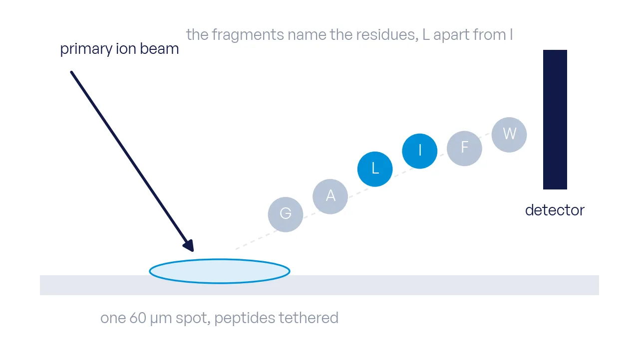
An ion beam hits one peptide spot on the array surface; lettered fragments fly off toward the detector, leucine and isoleucine as two distinct pieces.

2023 · International Journal of Molecular Sciences

DOI

Label-Free Imaging of Solid-Phase Peptide Synthesis Products and Their Modifications Tethered in Microspots Using Time-of-Flight Secondary Ion Mass Spectrometry

Time-of-flight secondary ion mass spectrometry reads the synthesis products in 60 µm array spots without a label: a fragment table per amino acid and protecting group, signals that grow linearly with residue count, and leucine told apart from isoleucine.

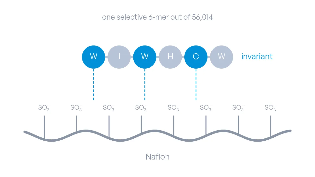
The Nafion chain with its sulfonate groups below, the 6-mer WIWHCW as six beads above, the three invariant residues in azure with contact lines to the chain.

2023 · Advanced Functional Materials

DOI

Selective Peptide Binders to the Perfluorinated Sulfonic Acid Ionomer Nafion

A screen of 56,014 random 6-mers against the fuel-cell ionomer Nafion finds the selective binder WIWHCW, locates its invariant residues by substitution and measures a dissociation constant of about 140 µM.

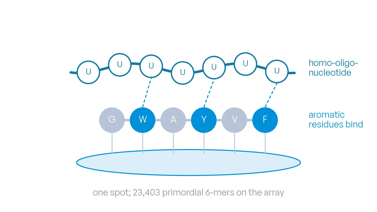
A short RNA strand of identical bases lies across a 6-mer peptide on a spot; the aromatic residues at the contact points are in azure.

2023 · Life

DOI

Screening for Primordial RNA–Peptide Interactions Using High-Density Peptide Arrays

Combinatorial libraries of primordial peptides, 23,403 6-mers, are screened against their cognate homo-oligonucleotides on high-density arrays to score which amino acids drive RNA–peptide binding.

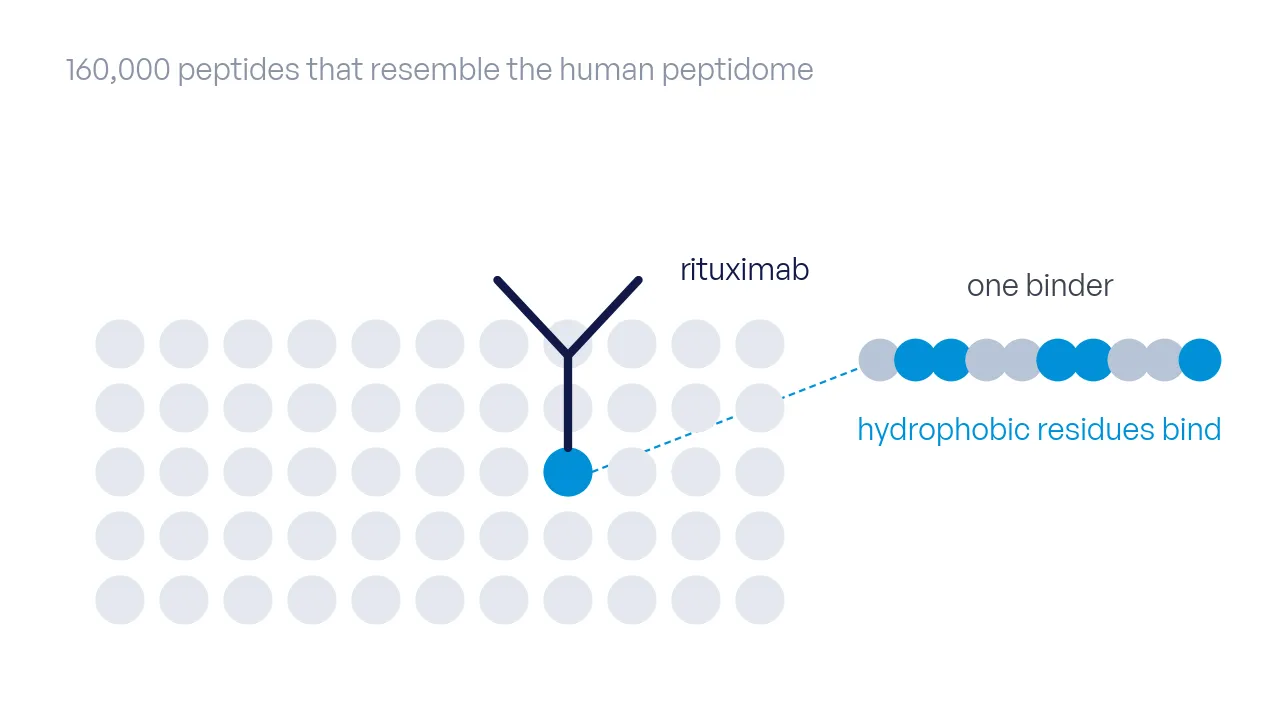
An antibody over a field of spots that stands for the human peptidome; one spot is held, and its peptide's hydrophobic residues are in azure.

2022 · International Journal of Molecular Sciences

DOI

Resemblance-Ranking Peptide Library to Screen for Binders to Antibodies on a Peptidomic Scale

A 160,000-peptide library ranked by resemblance to the human peptidome is screened for binders to the therapeutic antibody rituximab; substitution analysis of the 30 strongest gives dissociation constants in the hundreds of nanomolar range and a hydrophobic epitope.

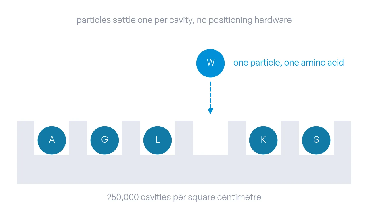
A row of microcavities in a surface; lettered amino-acid particles have settled one per cavity, one is still falling, and an arrow marks the coupling below.

2019 · Scientific Reports

DOI

Stochastic deposition of amino acids into microcavities via microparticles

Monodisperse microparticles loaded with amino acids settle stochastically into microcavities, one particle per cavity, and release their monomer for coupling: a route to molecular arrays at 250,000 spots per square centimeter without positioning hardware.

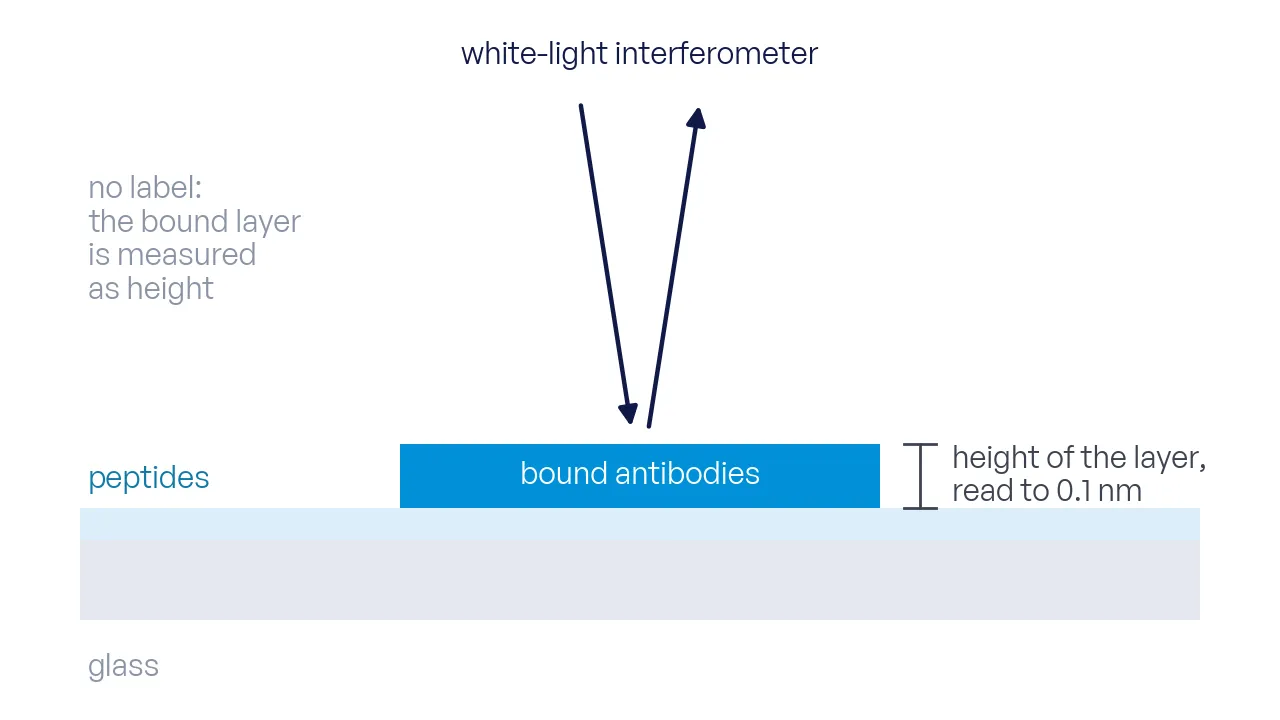
A spot in profile: the peptide layer on the surface, the bound antibody layer as a raised plateau, an interferometer beam from above measuring the height step.

2019 · High-Throughput

DOI

Vertical Scanning Interferometry for Label-Free Detection of Peptide-Antibody Interactions

Vertical scanning interferometry images antibody layers on peptide array spots without a label, at 1.4 µm lateral and 0.1 nm vertical resolution, and agrees with atomic force microscopy.

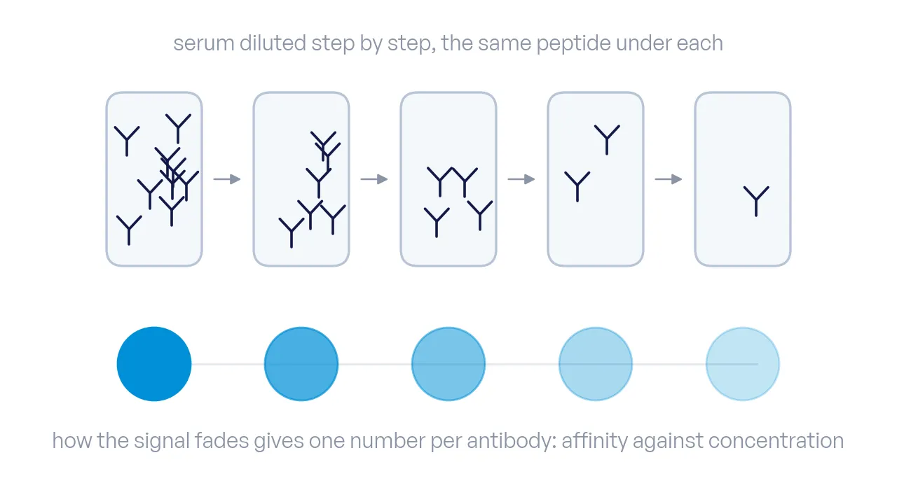
Five serum tubes in a dilution series with thinning antibodies; under each, the spot signal shrinks, and one line through the signals marks the serological number.

2019 · International Journal of Molecular Sciences

DOI

Serological Number for Characterization of Circulating Antibodies

Proposes the serological number, a dimensionless coefficient linking the dissociation constant of circulating antibodies to their serum concentration, and measures it on a peptide array with 15 motif peptides across serum dilutions.