
Epitope Mapping.
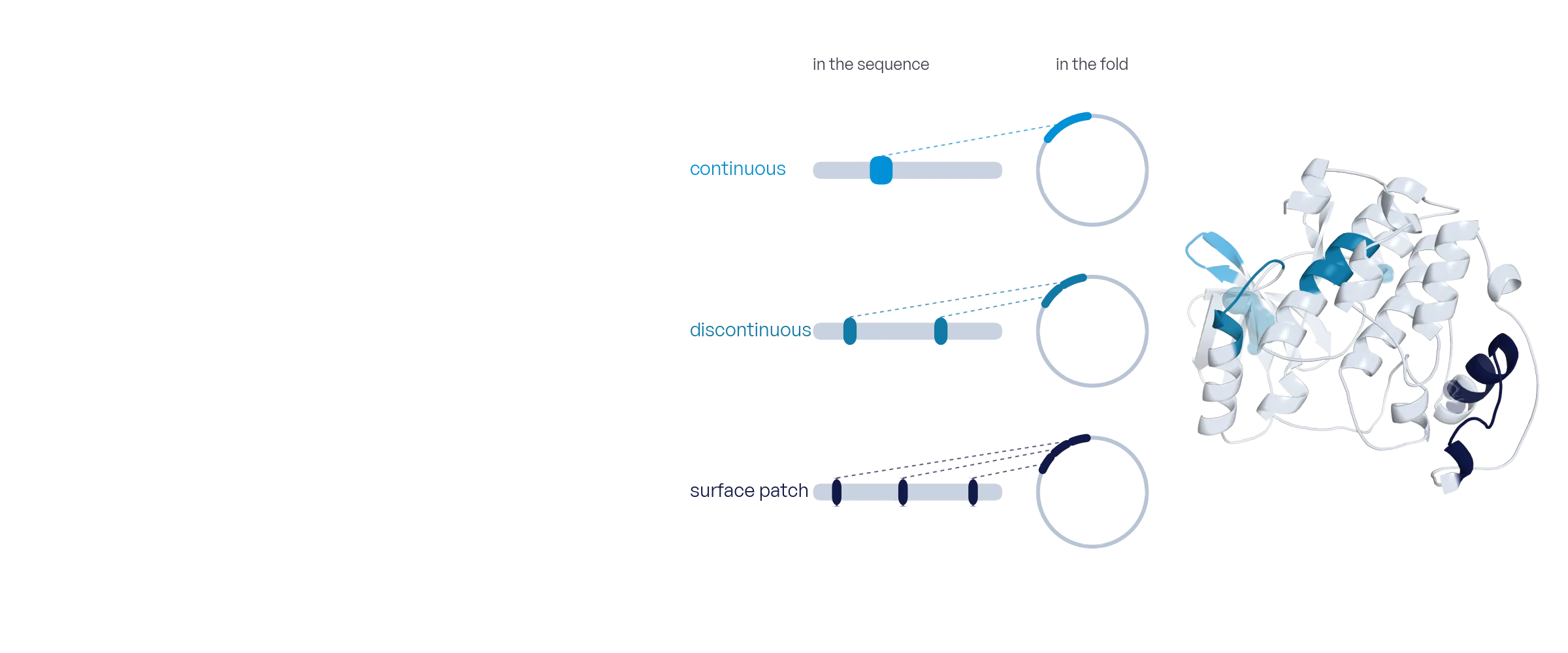
Continuous and discontinuous epitope candidates, derived from the experimental or predicted structure of your protein, tested on one peptide microarray. Structure-based candidates are unique to Axxelera.
What it answers
Where on the target protein does my antibody bind? Most epitopes are discontinuous, often called conformational: two or more stretches of the sequence, far apart in the chain, that fold into one contiguous surface patch. Conventional peptide libraries scan the sequence and overlook them.
We derive epitope candidates from the protein structure, an experimental one or an AlphaFold model: overlapping peptides along the sequence for continuous epitopes, and peptides that join two fragments or a whole surface patch for discontinuous ones. All candidates are synthesized on one microarray and probed with your antibodies, one per well. The result is the epitope map on the structure, with the confidence of each call.
When to use it
Antibody characterization
Locate the epitope of a monoclonal, continuous or discontinuous, bin a panel by the epitopes its members share, and document it for the filing.
Protein–protein binding sites
Probe a binding partner instead of an antibody and find the surface stretches that carry the interaction.
Serum and autoantibody profiling
Screen sera against candidates from a whole panel of antigens, up to 18 samples on one microarray.
Cross-reactivity
Test one antibody against candidates from related proteins, isoforms or species, side by side on one microarray.
How the assay works
Day 0
1Project submission
Name the target protein and send the antibodies. The candidate library is derived from the protein structure by us.
Day 1–7
2Peptide synthesis
The candidates are synthesized in situ on the microarray, cyclized where a candidate joins two fragments, along with blank spots and synthesis controls.
Day 8–10
3Bioassay
Each antibody, serum or binding partner is incubated in its own well, up to 18 on one microarray, next to a buffer well as reference. The microarray is scanned before and after.
Day 11–13
4Data analysis
Every candidate is tested against the background. Overlapping hits are merged into continuous epitopes with a core, fragment pairs and surface patches into discontinuous ones, and all are mapped onto the structure.
Day 14
5Project release
Raw data, processed results and the project report are released to you.
What you provide
Antibodies
Purified antibodies, sera or a binding partner, one per well. Detected with a labeled secondary antibody, or labeled directly. Amount and buffer are confirmed in the quote.
Target protein
The target as a UniProt entry, a sequence or a structure. Continuous candidates come from an overlapping scan of the sequence, discontinuous candidates from the structure, or send your own candidate list. Up to 25 residues per peptide.
Assay conditions
Optional. Preferred buffer, concentration and controls, such as an isotype or a pre-immune sample. Otherwise we propose them in the quote.
What we deliver
Raw data
Scanner exports and extracted signals for every spot and every well.
Processed results
Peptide summary and epitope tables: continuous epitopes with their core residues, discontinuous contacts and surface patches, and a specificity table across all wells, with z-score and false-discovery rate for every peptide. Spreadsheet and CSV.
Project report
Design, protocol, quality control, the epitopes mapped onto the structure with an interactive 3D viewer per antibody, and a data dictionary.
From a real project
Where do eleven antibodies bind two kinases?
- Question
- A diagnostics customer needed the epitopes of eleven monoclonal antibodies against two kinases, to bin the panel.
- Design
- For each kinase, overlapping 15-residue peptides along the sequence plus two-fragment and surface-patch candidates from the structure: 1,966 and 4,607 peptides. Two six-well microarrays, one antibody per well.
- Result
- Five of six antibodies against the first kinase gave an epitope. Four converge on one eight-residue stretch near the N-terminus and one binds a C-terminal stretch; several also bound two-fragment candidates, discontinuous epitopes a linear scan misses. One is conformational only. The five against the second kinase mapped no epitope; the control peptides were bright, so the negative is valid.
| Antibody | Epitope | Residues | Type | z-score | Class |
|---|---|---|---|---|---|
| AB-01 | core TGQ | 22–29 | continuous | 84.0 | epitope |
| AB-01 | two fragments | 55–62 · 109–115 | discontinuous | 35.3 | epitope |
| AB-02 | core TGQ | 22–29 | continuous | 75.8 | epitope |
| AB-04 | C-terminal | 282–294 | continuous | 13.7 | epitope |
| AB-05 | core TGQ | 22–29 | continuous | 61.2 | epitope |
| AB-05 | two fragments | 24–30 · 141–147 | discontinuous | 22.9 | epitope |
| AB-06 | — | — | conformational | 1.2 | not mapped |
Excerpt of the results table. Generic records, real columns.
Questions
How much antibody do I need for Epitope Mapping?
Send at least 20 µg of each antibody, or 50 µg if sample is not limited. The sample calculator in Learn (step 2.6) shows the amount per well for any concentration, and the quote confirms the one for your project.
What if the antibody needs the folded protein?
Some antibodies bind an extended surface that no peptide reproduces, and the map stays empty for them. The result is then a validated negative, with the control peptides bright, and we say so. It is worth knowing before a crystallography campaign.
Can you map sera or polyclonal samples?
Yes. Sera and pooled samples are run like antibodies, one per well, and the map lists every candidate the sample recognizes across the whole antigen panel. Individual donors need individual wells.
How many antibodies fit on one microarray?
One per well, up to 18 on one microarray, each seeing the identical candidate library, plus a buffer well as reference. Larger panels span several microarrays.
Can a peptide array map a discontinuous (conformational) epitope?
Yes, if the library is built from the structure. A linear scan tiles the sequence, so stretches that lie far apart in the chain never meet on one peptide. Candidates derived from the structure join them, and a hit names the fragments the antibody needs, drawn on the protein. An epitope that depends on the exact fold is better mapped by HDX-MS or a co-crystal structure.
Linear scan or structure-derived library?
Both, on one microarray. The overlapping scan finds continuous epitopes. The structure-derived candidates find the discontinuous epitopes that a scan overlooks: two fragments or a surface patch joined in one peptide, and cyclized to hold the shape.
Which structure do you use?
An experimental structure from the Protein Data Bank when there is one, otherwise an AlphaFold model. You can also restrict the mapping to a domain or an extracellular region.
Bring your target and your question
We will propose a library, a layout and a timeline within one business day.