
Protease Profiling.
Your peptides exposed to plasma or a protease on the microarray. A cleavage rate and a half-life for each peptide, and the cleavage motifs behind them.
What it answers
How stable is each of my peptides against proteases? We synthesize the library in situ, up to 15,000 peptides per microarray in a six-well layout, each peptide carrying a reporter that is lost when the peptide is cut, and expose the wells to your condition: plasma, serum, a purified protease, or a buffer at a chosen pH.
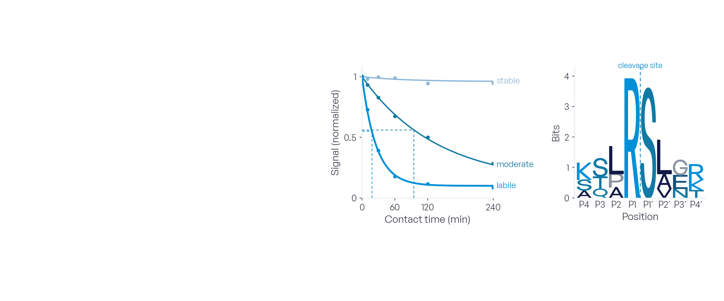
The wells carry the variable. Vary the contact time and every peptide gets a cleavage rate and a half-life. Vary the protease concentration at one contact time and every peptide gets an apparent EC50, the concentration that cleaves half of it. The result is a ranked table either way, with a stability class per peptide and the cleavage motifs derived from the data.
When to use it
Plasma stability
Rank thousands of therapeutic candidates by their half-life in plasma, and see which residues and modifications protect them.
Substrate specificity
Profile a purified protease against a substitution library around its cleavage site and derive its specificity motif from the data.
Counterscreens
Expose the same library to a panel of proteases, one per well, and find substrates cleaved by one and spared by the others.
Match the compartment
Run the assay where your peptide has to survive or be cleaved: plasma or serum, the acidic pH of an endosome or a tumor microenvironment, intestinal conditions. Up to 18 conditions side by side on one microarray.
How the assay works
Day 0
1Project submission
Configure the library and submit the project by email, with the protease or medium and the variable across the wells: contact times or protease concentrations.
Day 1–7
2Peptide synthesis
Your peptides are synthesized in situ on the microarray with a terminal reporter: up to 15,000 peptides in six wells, along with reference spots and control substrates.
Day 8–10
3Protease assay
Each well receives its own variable at 37 °C: its contact time, its protease concentration, or its own medium and pH. One well receives assay buffer only and is the null. One scan after the exposure measures the reporter that is left.
Day 11–13
4Data analysis
The signal loss of every peptide is measured against the null well and fitted. A contact-time series gives the cleavage rate and the half-life, a protease titration gives the apparent EC50, each with a confidence interval and a stability class. Comparing cleaved and uncleaved peptides gives the cleavage motifs.
Day 14
5Project release
Raw data, processed results and the project report are released to you.
What you provide
Sequences
A list of substrate sequences, including custom building blocks: unnatural or D-amino acids, cyclization by thioether, disulfide or amide, so that stabilizing modifications are tested in the same run. Or a substitution library we derive around a cleavage site. Up to 25 residues per peptide.
Protease or medium
A purified protease with its buffer, plasma or serum. Send your own, or we source it. Enzyme concentrations, pH and contact times are confirmed in the quote.
Assay conditions
Optional. Known substrates and non-substrates as benchmarks, preferred time points and temperature. Otherwise we propose them in the quote.
What we deliver
Raw data
Scanner exports and extracted signals for every spot at every contact time or condition.
Processed results
Stability summary and fitted curves: cleavage rate and half-life, or apparent EC50, each with a confidence interval, plus fraction lost, stability class and rank for every peptide, and the signal in every well. Spreadsheet and CSV.
Project report
Design, protocol, quality control, the ranking with figures, the cleavage motifs as sequence logos, condition comparisons where the design allows, and a data dictionary.
From a real project
Which of 7,494 candidates survive in plasma?
- Question
- A drug-discovery customer needed the plasma stability of a design library before choosing candidates for synthesis.
- Design
- 7,494 peptides of 10 to 20 residues, 6,206 linear and 1,288 disulfide-cyclized, with unnatural residues such as norleucine and N-methyl-leucine, in duplicate on one microarray. Six wells, one plasma contact time each, from 0 to 240 minutes at 37 °C, in pooled human plasma.
- Result
- 35 % of the peptides were cleaved within four hours, each with a half-life and confidence interval, and 65 % were stable. Susceptibility rose from 15 % for 10-residue peptides to 38 % for 18-residue ones, and the residue odds ratios showed which building blocks protect and which expose.
| Rank | Peptide | Form | Half-life | 95 % CI | Loss at 240 min | Class |
|---|---|---|---|---|---|---|
| 1 | PEP-03118 | linear | < 10 min | — | 82 % | fast |
| 2 | PEP-00472 | linear | 14 min | 9–21 | 79 % | fast |
| 3 | PEP-05930 | cyclic | 26 min | 18–37 | 74 % | fast |
| … | ||||||
| 974 | PEP-02207 | cyclic | 215 min | 160–290 | 38 % | slow |
| — | PEP-01144 | cyclic | > 240 min | — | 6 % | stable |
Excerpt of the results table. Generic records, real columns.
Questions
Can the library contain unnatural amino acids or cyclic peptides?
Yes. Any commercially available Fmoc-protected amino acid with a free carboxyl group, sourced per project, including D-amino acids. Cyclization by thioether, disulfide or amide. Up to 25 residues. Stabilizing modifications are tested in the same run as the unmodified sequence.
Plasma, buffer or a purified protease?
All three. A purified protease in its buffer gives its specificity motif. Plasma or serum gives the stability that matters for a therapeutic candidate. Buffer series across pH show where an enzyme is active, and let you emulate the compartment your peptide must survive. One microarray holds up to 18 conditions.
How is cleavage measured on a microarray?
Every peptide carries a reporter at its free end. When a protease cuts the peptide, the reporter is released and the spot loses its signal. One well receives assay buffer only and is the null: each treated well is measured against it, and reference spots inside every well correct for well-to-well differences. One scan after the exposure is enough.
Half-life or apparent EC50?
Both are available, and the wells decide which. Vary the contact time and the fit gives a cleavage rate and a half-life per peptide: the question is how long a peptide survives. Vary the protease concentration at one contact time and the fit gives an apparent EC50, the concentration that cleaves half of it: the question is how little enzyme it takes. Both are rankings on a surface, not solution constants.
How are the cleavage motifs derived?
By comparing the sequences of cleaved and uncleaved peptides position by position, as a sequence logo. The assay ranks substrates and derives the motifs from the ranking. It does not localize the cut site within a peptide.
Is the half-life absolute?
No, and neither is the apparent EC50. The peptide is tethered to the surface, so both are rankings under the chosen condition, not solution constants. Overlapping confidence intervals are not ordered. Confirm the leads in solution, for example by LC-MS.
How does on-array Protease Profiling compare with an LC-MS stability assay?
They fit different stages. LC-MS follows the intact peptide and its fragments in solution, so it gives a solution half-life and the cut site. It is the better choice for the last few candidates. The array compares up to 15,000 peptides on one microarray, all in the same plasma or protease, and ranks them with the motifs behind the ranking. On the array the C-terminus is anchored, so carboxypeptidases cannot reach it; in solution they can. The array narrows the library; LC-MS confirms the leads.
Bring your target and your question
We will propose a library, a layout and a timeline within one business day.